Trichodectes canis
Life History: T. canis are an ectoparasite visible with the naked eye. Trichodectes spp. have been found on carnivores and ungulates world wide. They are Chewing Lice (Malophaga) that are dorsoventrally flattened. They are not as well equipped to take in blood meals as sucking lice are but will imbibe blood meals, host fur, or sloughed skin. Female lice glue eggs to the hair shafts. Eggs will often remain attached even after the lice have hatched and will remain in place until the hair is shed. Average life span of the lice is ~30 days. Mating occurs on the host. The most common method of movement from one host to another is close contact. Typically lice are very host species specific but may infect other closely related species. They live their entire lives on the host. In most cases lice have coevolved with their hosts.
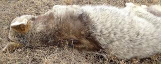
Clinical Signs: Varying degrees of alopecia (hair loss), broken guard hairs, matted underfur and skin lesions will be seen. Often scratching and irritation lead to secondary bacterial infections.
Intermediate hosts: T. canis can serve as an intermediate host to the tapeworm Diplyidium caninum.
Diagnosis: To find lice on live animals or freshly dead animals part the hair and look at the base of the hair shafts. Once the host is dead, lice will be gone from the host within 24-48 hours.
Management Implications: Usually there are little to no management implications at the population level since the lice typically infest hosts at low numbers and the hosts are most often immunocompitent. Instead the presence of lice can be an indicator of population dynamics. Since close contact is needed for the lice to move from host to host an increase in prevalence is an indication of high numbers of susceptible hosts in a given area. Fur or pelts of infested individuals may be downgraded because of decreased quality.
Zoonotic Potential: Since the lice are very species specific there is little to no zoonotic potential.
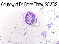
Trichodectes canis recovered from coyote skin (Figure 2, 3 A and B, 4 Courtesy of Dr. Betsy Elsmo, SCWDS)
Figure 1: Left: T. canis egg or “nit” on hair shaft. Right: Adult T. canis louse, approximately 0.7 mm long.
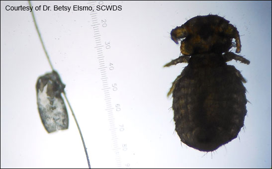
Figure 2: A and B: Adult T.canis lice in histologic section.
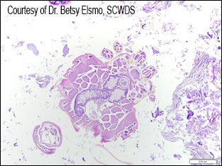
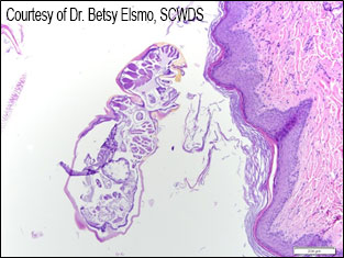
Figure 3: T. canis egg or “nit” in histologic section.
